
GEORGIAN RADIOLOGY CONSULTANTS
Medical Imaging is convenient, fast and easy.

Review: Bone Mineral Densitometry
Normal Bone
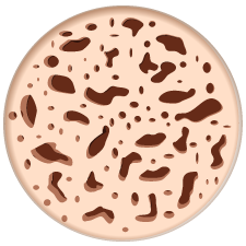
Osteoporosis
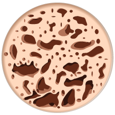
Severe Osteoporosis

Bone Mineral Densitometry (BMD)
What is Bone Mineral Densitometry?
Osteoporosis is a disease characterized by low bone mass and deterioration of bone tissue. This leads to increased bone fragility and risk of fracture, particularly of the hip, spine and wrist. Osteoporosis is often known as “the silent thief” because bone loss occurs without symptoms. Bone densitometry measures the bone mineral content in various sites of the body, allowing a physician to diagnose osteoporosis and assess a patient’s risk of suffering bone fractures.
Bone Health Assessment
Bone health assessments are targeted towards patients under investigation for vertebral compression fracture. We offer convenient and comprehensive appointments including nuclear medicine bone scan, x-rays, and/or densitometry to offer a holistic picture of your patient’s bone health, in one appointment. The assessment may be followed by a later consultation for vertebroplasty with our Interventional Radiologists if desired.
Use our Bone Health Assessment Requisition Form to streamline the process from diagnosis to treatment.
Nuclear Medicine
We are offering nuclear cardiac services such as Myocardial Perfusion Imaging (MPI) with pharmacological (Persantine) stressing and MUGAs .
We are also offering an array of non-cardiac nuclear diagnostic imaging namely Bone Flow and Scan (specific site), Total Body Bone scan, SPECT, Hepatobiliary Flow and Scan, RBC Liver Scan, Liver/Spleen Flow and Scan, Renal Flow and Scan, Salivary Scan, Parathyroid Scan, Perfusion Brain Scan.
Review: Integrated Prenatal Screening (IPS)
Integrated Prenatal Screening (IPS)
Screening Recommendations
The Society of Gynecology and Obstetrics of Canada recommends that “all pregnant women in Canada, regardless of age, should be offered, through an informed consent process, a prenatal screening test for the most common clinically significant fetal aneuploidies in addition to a second trimester ultrasound for dating, growth, and anomalies.”
The most common method of fulfilling this is to do an Integrated Prenatal Screening (IPS) testing combined with an 18-20-week ultrasound.

Integrated Prenatal screening is a three-step process.
- Nuchal translucency (NT) and early anatomy ultrasound. This is done at 11-13 weeks, but ideally should be scheduled between 11 weeks 5 days and 13 weeks 6 days. The gestational age is confirmed. The tissue at the back of the fetus’s neck (NT) is measured. (Babies with trisomy 21 and other aneuploidies tend to accumulate more fluid here)
- The first blood test must be done on the same day. Your patient must have the correct paperwork with them to complete the test. This test measures proteins in the blood and is used to modify the risk rating.
- The second blood test is done between 15 – 18 weeks.
Results
Results are labeled as either screen positive or negative.
- Screen Positive – If the results indicate a greater than 1/200 risk of Down Syndrome or a 1/100 risk of Trisomy 18 or 13 they are considered positive. 2% of all tests will be positive. Most will be false positive. Close to 90% of the pregnancies affected with Down Syndrome will screen positive (true positive).
- Screen Negative – A screen negative result indicates the chance of Down Syndrome is less than 1/200. More than 99% of pregnancies with normal 21, 18 and 13 chromosomes will screen negative. Please remember that 10% of pregnancies with Down Syndrome will also screen negative (false negative).
Other Benefits
- Placental Function – The same biochemical factors that are used in IPS testing have been found to reflect placental function and can identity a group of patients at risk for placental insufficiency. These patients have higher rates of IUGR, SGA, premature delivery and fetal demise. This group of patients can be further stratified by examining the maternal uterine arteries.
The IPS is considered to be false positive when both aneuploidy (trisomy 21, 18 and 13) and neural tube defects are excluded. Adverse pregnancy outcomes have been associated with false positive IPS with the following abnormal maternal serum markers:
1st trimester 10-13 weeks blood work:
low papp-a (<0.35 MOM)
hCG >5.0 MOM
inhibin A >3.0 MOM
2nd trimester 15-20 weeks blood work:
AFP >2.0 MOM
In the setting of a false positive IPS described above, measurement of maternal uterine artery Doppler has been shown to identify a group of pregnant women with the highest risk of adverse pregnancy outcomes. Uterine artery Doppler is typically performed between 18-22 weeks and an abnormal Doppler measurement with the maternal uterine artery Pulsatility Index (PI) > 1.45 suggests there is high risk for subsequent placental insufficiency. This patient will require early referral to an obstetrician. A normal PI is reassuring but should be followed with a third trimester ultrasound for fetal growth.
We ask that if you receive an abnormal IPS result, proceed with the 18 to 20-week ultrasound already booked, refer to genetic counselling for further testing, and refer the patient for uterine artery Doppler (18-22 weeks). In Barrie, this test is currently offered at the RVH but services will expand to the local clinics if demand is high. Please fax the IPS results with the ultrasound requisition.
Uterine Artery Doppler should also be considered in women at risk of placental pathology with current obstetric risk factors (pre-existing hypertension, gestational hypertension, thrombophilia, pre-existing renal disease and Type 1 Diabetes with vascular complications) as well as past obstetric risk factors (early onset gestational hypertension, placental abruption, IUGR and stillbirth).
Uterine Doppler will not be done in low risk pregnancies as it is not proven as a useful tool.
- Twin Pregnancy – If your patient is having twins, the chronicity can be most accurately assessed at an early ultrasound. This will affect your patient’s management throughout the pregnancy. Identical twins are at higher risk of complications. Monochorionic/monoamniotic and monochorionic/diamniotic pregnancies need early referral to obstetrics. Blood tests are not accurate in twins or triplets so the best screening test is the nuchal translucency which is unique to each baby.
Useful Links
The Society of Obstetrics and Gynecology of Canada 2007 Guidelines on Screening for aneuploidy: http://www.sogc.org/media/pdf/advisories/JOGC-feb_07-CPG.pdf
Mt Sinai IPS website: http://www.mountsinai.on.ca/care/pdmg/tests/ips
Screening Guide for Patients: Click here to download
Reference Guide for Health Care Providers: Click here to download
Review: Thyroid Nodules
Thyroid Nodules
Epidemiology
Thyroid nodules are extremely common in the population. Approximately 50% of the adult population have thyroid nodules on ultrasound. Fortunately, only 3-7% of these nodules are malignant.
Nodules are more common in:
- Elderly persons
- Women
- Iodine deficiency
- History of radiation exposure

Risk factors for carcinoma include:
- Age less than 20 years or more than 60 years
- History of neck irradiation
- Family history of thyroid cancer
- Male sex
- Previous diagnosis of type 2 MEN
Incidence of Thyroid cancer has increased over the last 30 years. The cause for this is uncertain but may be related to:
- Better diagnosis
- More FNA with US guidance (increased accuracy)
- Rise in radiation exposure in the general public
Ultrasound
Management
The decision to biopsy is based on the patient’s risk factors, the size and appearance of the nodule and the presence of suspicious cervical lymph nodes. In general, only solid nodules larger than 10mm should be considered for biopsy. Other nodules should be followed by ultrasound in 6-12 months and regularly thereafter. If a nodule is thought to be enlarging, then follow-up can be performed sooner.
Useful Links
The 2009 American Thyroid Association Guidelines for Management of Thyroid Nodules and Differentiated Thyroid Cancer: Progress on the Road from Consensus- to Evidence-Based Practice are available through this link: http://www.liebertonline.com/doi/pdf/10.1089/thy.2009.1601
American Association of Clinical Endocrinologists Medical Guidelines for Clinical Practice for the diagnosis and management of Thyroid Nodules. 2006. https://www.aace.com/files/thyroid-guidelines.pdf
Requisition Form
Before coming to one of our clinics, please remember:
To view the form, you will need Adobe Reader. You may download Adobe Reader for free at www.adobe.com. Please bring your completed form with you to our office at the time of your visit.
Book Your Ultrasound, BMD, Nuclear Medicine Appointment
To book an appt, please FAX the requisition to 705-726-8056
Or for your Ultrasound, BMD, Nuclear Medicine Appointment BOOK HERE with your name, phone number, and a clear copy of your requisition.
X rays are walk in only. Please do not email/fax X ray requisitions.
Things To Remember
Before coming to one of our clinics, please remember:
1. Your valid health card.
2. A signed requisition order or high-risk requisition order from your doctor.
3. Wearing masks is optional but encouraged.
View Our Articles
Find out more about our services by reading reading our blogs below.

Visit Georgian Radiology’s new location in Little Lake
Georgian Radiology is pleased to announce the opening of our newest clinic in the Little Lake Health Centre on June 22, 2015. This new location will allow us to expand our services in order to

Georgian Radiology
In Ontario, very few, if any medical practices are truly full-service – in other words, able to provide anything and everything that any one patient might require, from ultrasound to stress testing to a bone